Retinal Detachment Surgery in India - Expert Ophthalmologists, Hospitals & Cost
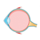
The retina is a part of the eye consisting of layers of light-sensitive nerve tissues and is present at the back of the eye. Its function is to receive the visual signals from the eye and transmit them as electrical signals to the brain through the optic nerve. Retinal detachment (RD) occurs when the retina pulls away from its normal position at the back of your eye. The retina becomes dysfunctional, which causes partial or complete loss of vision depending on the extent of the retinal detachment disease. When this happens, it is considered a medical emergency that requires retinal detachment surgery because when your retina becomes detached, its cells are severely deprived of oxygen, which may lead to irreversible blindness. You should not wait until you feel any pain because this condition is usually painless. The detached retina operation should be started as soon as possible.
Although retinal detachment disease is an ocular emergency that causes significant vision loss, it is a treatable condition that requires specialised medical care. However, because of inadequate medical facilities for the treatment of RD in developing countries, the risk of developing blindness caused by RD becomes relatively higher. In addition to this, many people with RD in developing countries have a late diagnosis of this condition. Studies show that about 69 percent of people have a one-month late diagnosis after the symptoms were experienced. Because of this, many people are unable to receive the treatment on time.
Seeking early diagnosis and treatment, many people from African nations like Ethiopia, Kenya, Tanzania, and Ghana travel to India for the treatment of retinal diseases. This is because the Indian eye clinics and hospitals have the latest technological medical equipment, which helps to provide early diagnosis, and the retinal detachment doctors in India are also highly experienced and known to provide the correct diagnosis and treatment. Among the many different countries that provide good quality medical treatment, India is the most chosen country for medical care by international patients as the treatments are of world-class quality, and along with state-of-the-art eye hospitals for retinal detatchment, it has the best retinal detachment surgeons and the cherry on top is the most affordable and profitable cost of the treatment. For patients researching the retinal detachment cost in India, CureIndia provides complete pricing transparency with internationally trained ophthalmologists, advanced surgical facilities, and all-inclusive packages at a fraction of the cost charged in the USA or UK.
What are the Symptoms of Retinal Detachment?
Most people do not experience any symptoms initially when the retinal detachment disease starts, and they develop the symptoms gradually as the disease progresses. The visibility of the retina detachment symptoms depends on the severity of the detached retina. If a small part of the retina is detached, you might not have any symptoms. However, if a large part of the retina gets detached, you are more likely to develop its symptoms. The common retina detachment symptoms include:
- Photopsia: you might suddenly start seeing flashes of light in the temporal part of the vision (away from the nose).
- The sudden appearance of small spots such as strings, flecks, lines, etc. floating before the eyes.
- Partial vision loss: it seems like a curtain is placed in front of your eyes and you are seeing a dark shadowing vision.
- Darkening of side vision, also known as peripheral vision.
- Blurred vision with shadowing effect.
What are the Retina Detachment Causes?
Retinal detachment occurs when the part of an eye known as the retina, which is responsible for collecting the signals from the eyes and sending them to the brain, pulls away or detaches from the back of an eye. The retina detachment causes can include injuries, inflammation, structural changes, or any damage that has affected the part of the eye where the retina is present. The risk factors for RD include ageing, previous eye surgery, family history of RD, nearsightedness, posterior vitreous detachment, diabetes, etc.
What are the Types of Retinal Detachments?
Depending upon how the retina gets detached from the back side of an eye, there are three major types of retinal detachment, which are explained below.

■ Rhegmatogenous RD:
It is the most common type of RD, which usually develops as a person ages. It occurs when a small hole or a tear develops in the retina. Due to this tear, the vitreous humour (a gel-like fluid produced by the eyes that nourishes the eyes) passes through it and gets collected behind the retina. As more and more fluid gets collected, it pushes the retina and hence detaches it from the back of an eye.
■ Tractional RD:
People who have diabetes are more likely to develop this type of RD. It occurs when high blood sugar levels damage the blood vessels present in the eyes and form scar tissue. When the scar tissue gets bigger, it tugs the retina and pulls it away from the back of an eye.
■ Exudative RD:
It is also known as serous RD. This is a type of RD that develops even when there is no formation of a tear or hole in the retina. The main cause of this type of RD is the leakage of blood vessels, which causes swelling or inflammation (uveitis) behind the eye, and fluid from the leaked blood vessels gets accumulated behind the retina, which then pushes the retina away from the supporting tissues.
What are the Diagnostic Tests for Retinal Detachment?
The diagnosis of the retinal detachment disease is made based on the patient’s symptoms, medical history, family history, and some diagnostic tests. A comprehensive and thorough eye exam will be conducted by your ophthalmologist according to the symptoms you are having. Following are the common tests needed to confirm the RD disease.
■ Dilated eye exam:
It is a diagnostic test in which the eyes of a patient are first dilated with eye drops, and a special medical tool known as an ophthalmoscope is used to examine the back structures of an eye, including the retina. Special attention is given to see if there is a hole or a tear in the retina and check for any signs of a detached retina.
■ Visual acuity test:
It is a test that is used to check the clarity and sharpness of a patient’s vision. A patient is asked to read the letters or to identify the objects by placing them at different distances.
■ Tonometry:
It is a test that is used to measure the intraocular pressure (IOP) in the eyes. If there is a fluid buildup in the eyes, then the patient will have elevated IOP, which can lead to the detachment of the retina.
■ Optical coherence tomography (OCT):
It is a type of imaging test that is a non-invasive approach to determining the cross-sectional images of the retina. The images help in the evaluation of the retina and other abnormalities that may have developed in the retinal layers.
Best Retinal Detachment Surgeons in India
The Indian healthcare sector has witnessed significant growth over the years. In the area of eye care, retina detachment surgery in India in particular has come out to be a specialised medical field that is focused on treating various retinal diseases such as retinal detachment, diabetic retinopathy, etc. Indian eye clinics and hospitals have the most experienced and skilled retinal detachment ophthalmologists who have contributed greatly to the healthcare sector of India. Listed below are some of the top doctors for retinal detachment surgery in India:

2. Dr. Parul Maheshwari Sharma


4. Dr. Sameer Kaushal

LET'S PLAN YOUR
TREATMENT IN INDIA
What is the Cost of Retinal Detachment Surgery in India?
On average, the retina operation cost in India can range from 720 USD to 840 USD. However, it can rise up to 1,500 USD, and it can even go as high as 3,000 USD. This is because the total cost depends on various factors such as the patient’s overall eye health, type of surgery needed, surgeons’ fees, chosen hospital and its location, diagnostic test cost, medication cost, travel cost, insurance coverage, etc. Patients who want to plan their medical travel in advance and need a personalised estimate of the retinal detachment cost in India can contact CureIndia's case managers, who factor in all clinical and logistical components before providing a transparent all-inclusive quote.The costs of different treatments for retinal detachment surgery in India are listed below:
| Treatment |
Cost |
Stay in India |
| Retinal Detachment Surgery in India |
$2,000 per eye |
7-10 days |
| Vireo Retinal Surgery in India |
$2000-$2500 per eye |
5-7 days |
What are the Types of Retinal Detachment Surgery in India?
The primary aim of retinal detachment surgery in India is to reattach the retina, restore its normal function, and prevent it from getting detached completely. There are several options for the treatment of retinal detachment in India; the common ones are explained below:

■ Laser photocoagulation:
It is a laser retinal detachment surgery in India that is used for the treatment of small holes and tears in the retina. It is a painless procedure in which the surgeon will numb the patient’s eye with the help of anaesthetic eye drops, and then a special laser will be used to target the retinal tear. Laser is used to create controlled burns, which will make the scar in the tissues. Now these scar tissues will act like a wall in the retina.
■ Cryotherapy:
It is also known as cryopexy therapy, which uses cold temperatures to form scar tissue. In this procedure, the surgeon will numb the patient’s eye by injecting anaesthesia around the affected eye and then position a freezing probe over the area where the retinal tear is present. By doing so, the scar tissue will form, which will seal the tear and help the retina reattach to the back of the eye.
■ Pneumatic retinopexy:
It is a procedure that involves the use of gas bubbles in the eyes to push the detached retina back and reattach at its place. This procedure is followed by the laser treatment or the cryotherapy to seal the retinal hole.
■ Scleral buckle surgery:
It is one of the most common types of retinal detachment surgery performed in India. In this procedure, the patient will be sedated under general anaesthesia, and the procedure will be initiated by treating the retinal tear with laser treatment or cryotherapy. Then the surgeon will place a small band made up of silicone on the outside of the sclera (white portion) of an eye. This band will stay in the eye permanently, and it will move the sclera towards the middle of the eye, which will help the retina to settle back at its place.
■ Vitrectomy:
This procedure is mostly used for the complicated case of retinal detachment. This procedure involves making a small incision in the sclera of an eye and the extraction of excess vitreous fluid that has gathered and caused retinal detachment. This procedure is then followed by photocoagulation or cryotherapy in order to close the retinal tear.
In scleral buckle surgery and vitrectomy, at the end of the procedures, intraocular gas is injected to replace the vitreous fluid and help the retina reattach. The type of treatment selection depends on the type, severity, and location of retinal detachment and the overall eye health of a patient.
How is Recovery After Retinal Detachment Surgery in India?
After the retinal detachment surgery is completed, you may feel a little discomfort initially, but it will go away as you heal. The complete retina detachment recovery will take about a few weeks. You will need to wear an eye patch to protect the treated eye for a few days. Your ophthalmologist will prescribe some medication and eye drops that you will need to apply in order to prevent swelling in the eyes and boost the healing process. Also, make sure to keep your head position as recommended by your doctor for the first few days after the surgery. Abstain from going to crowded places, doing heavy exercises, doing any strenuous activities, etc. up until your doctor allows you to. Your eye doctor will tell you when you can resume your regular life activities.
The retinal detachment disease is an eye disease in which the retina, which is a light-sensitive layer of tissue present at the back of an eye, is detached and pulled away from its normal position. This can happen due to many reasons, such as an injury, increased eye intraocular pressure, etc. Initially, it does not show any symptoms, and the symptoms gradually appear as the disease progresses, which include blurred vision, seeing floaters, etc. If it is not treated on time, it can lead to loss of vision. While the detached retina operation is widely available in developed countries, developing countries are still struggling to provide its treatment because of insufficient medical resources and inadequate healthcare systems.
That is why a lot of people travel to other countries for the treatment. For example, a lot of people travel to India every year for different medical treatments, including the treatment of retinal detachment disease because India provides world-class treatment at a very low cost. The expertise of ophthalmologists, the most advanced hospital infrastructure, and amazing facilities make India a medical hub for international patients. Patients who have compared the retinal detachment cost in india against equivalent treatment prices in the USA, UK, or Australia consistently find that India delivers the same internationally certified surgical outcomes at savings of 60–70%, making it the most financially viable and clinically sound destination for retinal detachment surgery.
LET'S PLAN YOUR
TREATMENT IN INDIA
Retina Detachment Surgery in India FAQs
There is no preventative measure for the retinal detachment disease that is caused by ageing. However, you can reduce the risk of developing it by getting regular eye checkups and avoiding eye injury by wearing safety gadgets while doing any activities that can damage the eye.
No, the detached retina cannot be cured on its own, and it requires a surgical procedure to reattach the retina and to further prevent it from being more detached.
No. The detached retina cannot be reattached with the eye drops. It can only be repaired with a detached retina operation. If not treated early, the disease can progress and lead to blindness.
The most common retina detachment causes in adults are eye injuries, diabetic retinopathy, uveitis, ageing, family history, severe nearsightedness, posterior vitreous detachment, etc.
The success rate of retinal detachment surgery in India is very high in India. According to the reports, about 90 to 95% of detached retina cases are successfully treated with only one surgery. Only a few complicated cases require additional medical procedures.
Your vision will be blurry for several days after the retinal detachment surgery in India. It will slowly improve as your eye heals. It can take about a few weeks to 3 months for the vision to improve after the surgery.
You will need to avoid activities that can put strain on the eyes. For the initial days after the retinal detachment surgery in India, you should also avoid strenuous activities like intense exercise, heavy lifting, etc. Make sure to attend regular follow-up appointments to monitor your overall eye health.
If left untreated, the cells of the retina will be separated from the layer of blood vessels, which supplies nourishment and oxygen to the eye. If the detached retina is not treated and left as it is for a longer time, then it can lead to permanent loss of vision in the affected eye.
The retinal detachment disease in children is known as paediatric retinal detachment (PRD). It is a very rare condition and only accounts for 3 to 7 percent of all retinal detachment cases.
Nearsightedness, also known as myopia, causes the retina to stretch and become prone to peripheral retinal tears. Also, the myopia degenerates the vitreous (gel-like fluid in the eyes), resulting in its collapse and separation from the retina. All these factors lead to the detachment of the retina from the back of an eye.
Retinal detachment can occur in both eyes simultaneously, or it can only occur in one eye at a time. It differs from patient to patient. If you have a detached retina in one eye, then you should consider getting your eyes checked regularly to monitor the progress of the disease in the already affected eye and to check whether the other eye is also affected or not.


 +918448560641