Quick Overview
- Procedure: Advanced glaucoma surgeries (like canaloplasty) and stem cell therapy (for regeneration)
- Cost in India: $3,000 – $5,000
- Hospital Stay: 1 day
- Stay in India: 7–10 days
- Recovery: 1–2 weeks
- Success Rate: High for vision preservation (90%+ in early-stage glaucoma)
- Preffered Cities: Chennai, Delhi, Bangalore, and Mumbai
Optic Nerve Treatment in India: Cost, Top Doctors & Hospitals
India has emerged as a global leader in providing cutting-edge solutions for optic nerve disorders, offering hope even in complex cases. From advanced pressure management to regenerative therapies, India provide the best optic nerve damage treatment to help you see a brighter future.
Eye nerve damage treatment in India combines world-class ophthalmologists with the latest diagnostic technology. Whether you are dealing with glaucoma or atrophy, you can access specialized care at a fraction of the cost in the UK or USA.
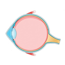
The optic nerve is a structure of the visual system, made up of nearly one million nerve fibers that transmit visual signals from the eyes to the brain. Each eye has its own optic nerve, which plays a crucial role in enabling clear and accurate vision. These nerve fibers originate from retinal ganglion cells and collectively form the pathway that allows the brain to interpret what we see.
At the point where the optic nerve exits the retina, there are no light-sensitive photoreceptor cells. This area creates a natural blind spot, which normally goes unnoticed because the brain seamlessly compensates for it.
One of the most common conditions affecting the optic nerve is glaucoma. Glaucoma occurs when intraocular pressure increases, leading to sustained compression of the optic nerve. Over time, this pressure damages the nerve fibers, causing progressive vision loss. This damage is referred to as optic nerve atrophy and, if left untreated, can lead to irreversible blindness.
India offers advanced and cost-effective optic nerve treatments, supported by experienced ophthalmologists, modern diagnostic tools, and internationally accredited hospitals, making it a preferred place for patients needing timely optic nerve treatment in India.
What is an Optic Nerve?
The normal role of the optic nerve is to transmit messages from the eye to the brain. In this capacity, the optic nerve acts as a messenger that assists in interpreting what is seen. It is the cornea's job to guide light entering the eye so that it may focus on the retina and produce the clearest picture possible. Light is seen by the retina, which then uses that information to generate impulses or currents. The current is carried through the optic nerve and delivered to the brain, which is responsible for interpreting it as a picture.

Even though the optic nerve's function seems to be rather straightforward, it is, in fact, an extremely important factor in our capacity to comprehend the world surrounding us. Because of how essential it is, researchers are hard at work developing a device that can perform the same functions as the optic nerve in the hope that this may allow patients who have suffered damage to the optic nerve to regain their eyesight.
In What Part of the Eye do we find the Optic Nerve?
One of the 12 cranial nerves, the visual, is the second. The optic nerves of each eye are distinct. The nerve that connects the eyes to the rest of the body:
- Begins in the optic disk, a collection of cells located in the retinal tissue in the rear of the eye.
- The optic canal is a bony hole in your skull via which light enters your brain.
- The optic chiasm is an X-shaped structure formed when the Optic nerve in one eye crosses over into the optic nerve in the other.
- The optical radiations are divided into upper and lower channels (ORs).
Messages sent by the OR pathways go to the visual cortex, a region of your brain. The visual cortex processes sensory data related to sight. There are a wide variety of conditions that may affect the optic nerve, including the following:
- Glaucoma is a collection of disorders that constitute the most common reason people in the United States become blind. Glaucoma is an eye condition that often develops due to a gradual increase in the fluid pressure within the eyes, which ultimately causes damage to the optic nerve.
- An atrophied optic nerve is called Optic nerve atrophy. Illness or trauma to hazardous chemicals may all contribute to the condition, as can insufficient blood supply to the eye.
- Swelling of the optic nerve is referred to as optic neuritis. There are situations when the root reason is unknown. Infections and diseases associated with the immune system, such as multiple sclerosis, can be the root cause.
- Over time, the accumulation of protein and calcium salts causes the optic nerve to develop pockets known as drusen in the optic nerve head.
Because of its structure, the Optic nerve is a sensitive marker that may be used to detect issues inside the brain. This nerve serves as a conduit between the retina and the head at the rear of each eye. The whole surface of the optic nerve is soaked in cerebral spinal fluid throughout its brief journey from the brain to the eye. This journey only takes a few millimeters.
What are Some Symptoms of Optic Nerve Damage?
Optic nerve compression happens when a mass develops in the brain, pushing on the optic nerve and causing eye pressure. Several different conditions may cause this. It may result in visual difficulties and perhaps blindness. Compression of the optic nerve may cause a variety of optic nerve symptoms, including impaired vision, which may also include:
- Hazy vision.
- A larger blind spot than before.
- The ability to see two things at the same time.
- Nausea and vomiting for no apparent reason.
- Reduced or eliminated eyesight in the periphery.
How is Diagnosis for Optic Nerve Damage Done?
Bright light will be utilised to examine the look of the head of the optic nerve and determine whether or not glaucomatous damage has occurred to the structure of the eye. The process takes very little time and causes no discomfort.
Slit-lamp examinations of the optic nerve include specialised lenses and often involve NO touch being made with the patient's eye. The examiner utilizes a portable instrument to view the eye's internal structures by positioning themselves close to the patient. A piece of equipment called a direct ophthalmoscope might also be used to perform examinations on the optic nerve and the optic disc.
An image of the optic nerve could also be obtained to maintain a record and monitor any shifts that may occur over time. For this test, the pupil will need to be dilated.
Experienced Surgeons for Optic Nerve Treatment in India
India is home to some of the leading ophthalmologists who specialise in optic nerve treatments, offering advanced diagnostics and personalised treatment plans. Among the leading experts in this field are Dr. Suraj Munjal, Dr. Suwarn Chetan, Dr. Sumit Gupta, and Dr. Priti Kumari, each known for their extensive expertise and successful track records. Let’s hear from some of our top optic nerve treatment doctors in India:
1. Dr. Suraj Munjal
Dr. Suraj Munjal is a a top eye doctor in India who is known for his expertise in diagnosing and managing optic nerve-related conditions. He specialises in providing advanced eye care, evaluation of optic nerve disorders, and personalised treatment planning. With experience of over 20 years, Dr. Munjal is one of the most preferred doctors for optic nerve treatment in India.
2. Dr. Suwarn Chetan
Dr. Suwarn Chetan is an experienced eye specialist in India dedicated to the treatment of various optic nerve conditions. His clinical practice focuses on early diagnosis, advanced imaging, and evidence-based management of optic nerve issues. Dr. Chetan is known for his meticulous evaluation and commitment to providing the highest standard of ophthalmic care.
3. Dr. Priti Kumari
Dr. Priti Kumari is an experienced ophthalmologist for optic nerve treatment in India with specialisation in evaluating and treating optic nerve conditions. She is known for her precision, comprehensive care strategies, and supportive patient communication.

4. Dr. Sumit Gupta
Dr. Sumit Gupta is a trusted name in ophthalmology, particularly in optic nerve disorders. He is trained in modern diagnostic techniques and offers comprehensive treatments. Dr. Gupta’s experience in handling diverse optic nerve cases makes him a preferred specialist for patients needing accurate assessment and long-term visual treatment.
What is the Cost of Optic Nerve Surgery in India?
Many individuals are traveling from African countries to India for Optic Nerve Surgery. The Indian Eye Hospitals have the most recent technologies and provide premium facilities and treatments. Also, the cost of Optic Nerve Surgery in India is lowest as compared to other countries. The Eye specialists in Indian hospitals have years of training and experience which is beneficial for providing the best medical service. The new approach for Optic Nerve Surgery which is Stem Cell Therapy is included in Indian eye hospitals. Listed below is the average cost of Optic Nerve Surgery in India.
| Treatment |
Cost in India (USD) |
Stay in India |
| Optic Nerve Surgery in India |
$3,000 to $5,000 |
7–10 days |
Comparison of Optic Nerve Surgery cost in US vs India
Optic nerve treatments in the USA or UK can be incredibly expensive, often ranging from $12,000 to $25,000 depending on the complexity. In India, the same level of expertise and technology costs between $3,000 and $5,000. This massive saving allows patients to access long-term follow-up care and rehabilitation services that might otherwise be unaffordable in their home countries.
| Location |
Cost (USD) |
| India |
$3,000 to $5,000 |
| USA |
$12,000 – $25,000 |
Optic Nerve and Intraocular Pressure
The increased pressure in the eyes gradually causes damage to the nerve fibers in the retina and the optic nerve, which results in a loss of vision. On average, the typical range for intraocular pressure is between 10 and 21 mmHg. At higher than 21 mmHg, retinal and optic nerve fibers may be progressively damaged, damaging the major optic nerve that carries visual impulses to one's brain. If it is not diagnosed promptly, it might lead to a loss of vision that cannot be reversed.

To further grasp this concept, think of your eye as a balloon inflated with water. If there is more water, the balloon will grow taut. Because of the microstructural changes that have taken place in certain regions, either less fluid is created or drained, causing an increase in pressure. Similarly, an increase in the pressure inside the eyes may affect the optic nerves located in the rear of the eye. Because of this, it is essential to keep the portion of the eye responsible for producing fluid in equilibrium with the area responsible for emptying that fluid.
What are the Options for Optic Nerve Damage Treatment in India?
Early indications of optic nerve injury, such as visual distortion and other issues with perception, may be modest. This is even though optic nerve injury is a significant neurological ailment triggered by an even more catastrophic fundamental disease or disorder. In most cases, vision loss is just temporary. In most cases, it gets better on its own over a few weeks or months, at which point therapy may not be necessary. Moreover, there is a possibility that the visual loss will be permanent in certain circumstances.
Physicians may prescribe steroid medication when the symptoms are very severe, for instance, when the condition afflicts both eyes. Steroids have been shown to hasten healing from optic neuritis; however, these medications do not affect how effectively the eyes heal. Nevertheless, using steroids for an extended period might result in negative side effects such as elevated blood sugar levels, increased body fat, and bone difficulties. Additional eye nerve treatment for Optic nerve damage that may be administered at home include the following:
- Abstaining from taking hot baths and engaging in strenuous activity
- Consuming meals that are good for you
- Avoidance of tobacco
- Consuming a great deal of water
Stem Cell Treatment in India for Patients with Optic Nerve Damage
The most comprehensive selection of stem cell treatment programs available for incredible improvements to one's health. Damage to the optic nerve causes a disruption in the normal transmission of visual information to central targets, which ultimately leads to the extinction of retinal ganglion cells and an irreversible loss of vision. Experiments have been done to see whether treatments using mesenchymal stem cells derived from various sources will improve the survival rate of retinal ganglion cells and promote their regeneration.
Stem cell therapy is currently one of the major paths for treating the nerve of the eye. This therapy includes pre-treatment thorough diagnosis in a hospital, specific selection of the appropriate cellular material administration by competent physicians, and all-around care and support throughout the monitoring and recovery process.
The ability of stem cells to undergo self-renewal and regeneration, cell division through mitosis, and differentiation into specialised cell types is the primary benefit of employing stem cells as a therapy method. These characteristics allow stem cells to be used in treating various organs and body systems.
What are Some Precautions to Take In Order to Protect the Optic Nerve?
Optic nerve damage is mostly caused by poor blood flow, which makes it difficult to supply nutrients and eliminate waste items, as well as oxidative stress, which damages and kills cells throughout the eye. There are methods available to fight back against this:
- Ensuring that the optic nerve continues to get a healthy amount of blood flow. Healthy vision relies on regular blood flow to the retina and optic nerve. The blood supplies vital nutrients and oxygen, which are essential for maintaining the optic nerve's health.
- Promoting the health of the mitochondria. A human cell's mitochondria are the cellular organelles responsible for energy production. They do this by absorbing nutrients from the blood and producing high-energy molecules for the cell. To maintain a healthy optic nerve, a person must have healthy mitochondria.
- Preserving a normal level of intraocular pressure. A normal eye pressure indicates that the optic nerve is in good condition. Maintaining a healthy optic nerve is essential by paying close attention to the pressure within the eye, also referred to as intraocular pressure.
- It is essential for the health of the optic nerve to restrict one's exposure to oxidation. The retinal cells, which are necessary for vision and send signals to the optic nerve, are susceptible to damage from oxidation. Using antioxidants reduces one's level of exposure to oxidation.
LET'S PLAN YOUR
TREATMENT IN INDIA
Optic Nerve Treatment in India FAQs
Optic nerve injury cannot be corrected since it is permanent once it has occurred. There are no nerve fibers in the optic nerve capable of self-regeneration. Thus they must be replaced. The body cannot repair broken nerve fibers.
As long as the optic nerve heals properly, most patients will regain all of their vision within the first several months.
Other health issues, one of the most common causes of optic neuritis is an infection caused by bacteria or viruses. Behcet's disease, Sarcoidosis, and lupus are all diseases that may induce recurrent ocular neuritis, as can other autoimmune diseases.
A slit light is used to examine the Optic nerve without touching the eyeball. The Direct Ophthalmoscope may also inspect the optic nerve or optical disc. By getting near the patient and using a handheld instrument, the examiner can see what's happening within the eye.
Effects that last for a long time, you may safely use castor oil on your eyes because of its antibacterial and anti-inflammatory characteristics. One further advantage of using eye drops made from castor oil is how prolonged they continue to be effective. It's been shown that they may remain in your eyes for up to four hours, according to research.
While traditional medicine focused only on "stopping further damage," modern treatments in India, including neuroprotective therapies and regenerative medicine, aim to improve the health of remaining nerve fibers. Early treatment offers the best chance for vision improvement.
Yes. Glaucoma treatment focuses on reducing eye pressure to prevent nerve damage. Optic atrophy treatment focuses on revitalizing the damaged nerve fibers and improving blood flow to the eye.
To provide you an accurate plan, we typically need your OCT (Optical Coherence Tomography) scan, visual field test (perimetry), and any recent MRI of the brain/orbits if available.
Not always. Many optic nerve conditions are managed with specialized injections, advanced medications, or non-invasive therapies. Surgery is usually reserved for cases like glaucoma, where pressure must be physically lowered.
Very safe. India is home to JCI-accredited eye hospitals (like Sankara Nethralaya or LVPEI) that are world-renowned. At CureIndia, we ensure a seamless journey, from airport pickup to your final follow-up, with total focus on your safety and comfort.


 +918448560641